Understanding Myocardial Infarction: Key Diagnostic Tests
"Doctors use a combination of electrical readings, blood chemistry, and visual imaging to determine if the heart muscle is actively dying or merely struggling for oxygen."
When you feel a crushing pressure in your chest, the medical team moves quickly to differentiate between stable angina and a life-threatening myocardial infarction. They primarily rely on electrocardiograms (ECG), specific cardiac enzyme blood tests, and advanced imaging to map the damage.
Key Takeaways * ECG (Electrocardiogram): The immediate first step to detect electrical irregularities, such as ST-segment changes, which classify the type of heart attack.
* Blood Tests: Measuring troponin levels (specifically high-sensitivity troponin) identifies actual damage to the heart muscle cells. * Imaging & Invasive Tests: Procedures like coronary angiography or CT scans allow doctors to see the physical location of arterial blockages.
* Clinical Context: Diagnosis depends on the interplay between physical symptoms, the timing of the pain, and the patient's specific risk factors.

Can an ECG help me diagnose a heart attack? At midnight in the dim emergency room, the technician presses cold, adhesive sensors against the patient's sweating skin.
It is 3:15 AM, and the fluorescent lights of the emergency department hum overhead as a technician places cold, adhesive sensors across a patient's chest and limbs.
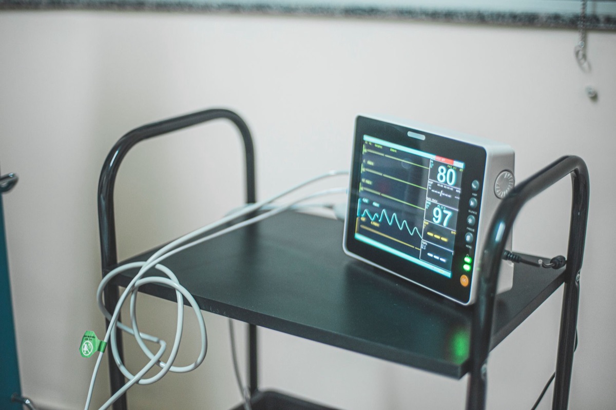
You watch the monitor, seeing the jagged green lines dance across the screen, representing the electrical rhythm of a heart under duress.
This 12-lead ECG is often the very first tool used to look inside the heart's electrical system. It serves as the frontline defense in identifying the specific nature of a cardiac event.
It allows clinicians to distinguish between a STEMI (ST-segment elevation myocardial infarction), which involves a complete blockage, and an NSTEMI (non-ST-segment elevation myocardial infarction).
Specific patterns, such as an elevation of the ST-segment by 1 mm or more in at least two contiguous leads, are strong indicators of acute myocardial injury.
Ischemic arrhythmias—irregular heartbeats caused by a lack of oxygen—are frequently associated with increased rates of illness and death, particularly during the first 48 hours after a patient is admitted to the hospital.
Because of this risk, the ECG is not just a one-time snapshot but often a continuous watch over the patient's electrical stability. But the electrical signal only tells half the story; the true damage often hides in the chemistry of the blood.
How do blood tests like troponin help in myocardial infarction diagnosis?
A nurse draws a vial of blood, the dark red liquid swirling in the tube as it is sent to the lab for urgent analysis. I remember sitting in a clinic waiting room once, watching a technician prepare a similar vial, realizing how much of our health relies on these tiny, invisible markers.
According to Health technology assessment (Winchester, England), high-sensitivity tests may offer better value for money than standard troponin tests that require repeat testing at 10–12 hours.
While the ECG looks at electricity, this blood test looks for the "debris" left behind by damaged cells. When heart muscle cells are injured or die, they leak specific proteins into the bloodstream. The most critical of these is troponin.
Typically, these levels begin to rise between 4 and 12 hours after the onset of the cardiac event, often reaching their peak concentration between 24 and 48 hours later.
Modern medicine has moved toward high-sensitivity troponin (hs-TnT) assays to speed up this process. These advanced tests are highly efficient for hospital resource management.
Health economic analyses have indicated that high-sensitivity tests may be considered value for money compared with standard troponin tests, which require repeat testing at 10–12 hour intervals.
While some older protocols still reference CK-MB (creatinine kinase-MB), troponin remains the gold standard because it is much more specific to the heart muscle. If the troponin levels are rising, it provides clear evidence that the myocardium is under direct attack.
However, even with clear blood chemistry, doctors often need to see the physical "plumbing" to fix the problem.
Which imaging tests can confirm my coronary artery disease? The patient lies still on a hard, cold table as a thin catheter is guided through the wrist or groin toward the heart. In the dim light of the catheterization lab, the medical team watches a high-definition monitor, waiting for the moment the dye reveals the truth.
This invasive procedure, known as coronary angiography, provides the most direct view of the coronary arteries. By injecting a specialized contrast dye, doctors can watch in real-time as the dye flows through the vessels.
If a blockage is present, the dye will stop or slow down, allowing the medical team to see exactly where a stent needs to be placed to restore blood flow.
For those who do not require immediate emergency intervention, non-invasive options like CT coronary angiography (CTCA) are available. This scan uses X-rays to create a detailed 3D map of the arteries, often detecting plaques or narrowing (stenosis) in about 10 to 15 minutes.
In certain clinical settings, such as preparing for major surgery, doctors may use cardiac functional examinations to assess risk.
For instance, in patients over 65 or those with known cardiac pathology, these functional exams help stratify the risk of developing cardiovascular complications during abdominal surgical interventions.
While imaging shows the structure, it doesn't always show how the heart behaves under pressure. That brings us to the necessity of physical exertion.

When are stress tests (exercise or nuclear) indicated?
A patient walks steadily on a treadmill, their breathing becoming heavy as the speed and incline increase. Sweat beads on their forehead, and the rhythmic thud of their footsteps matches the rising tempo of the machine.
According to Kardiologiia, cardiac functional examination methods are used for risk stratification in patients over 65 years of age or those with cardiac pathology.
An exercise stress test (often called an Ergo-test) monitors the heart's electrical activity via a 12-lead ECG while the patient reaches a target heart rate. If the heart muscle doesn't receive enough oxygen during this "stress," the ECG will show signs of ischemia (lack of blood flow).
If a patient cannot walk on a treadmill, a nuclear stress test may be used. This involves injecting a small amount of a radioactive tracer that travels through the blood.
A specialized camera then maps the blood flow to the heart, providing a visual representation of how well the tissue is being perfused over a 60 to 90-minute imaging period.
It is important to note that not all heart attacks are caused by a sudden blockage of a plaque. A condition known as type 2 myocardial infarction (T2MI) can occur when there is an increased demand for oxygen or a reduced supply, even without an acute blockage.
According to the Journal of the American College of Cardiology, type 2 myocardial infarction (T2MI) can occur due to increased myocardial oxygen demand or reduced supply even without acute atherothrombotic plaque disruption.
In these cases, stress testing may be a necessary step to rule out underlying ischemia.
What are the steps in the diagnostic workflow for angina or MI?
The clock starts ticking the moment a patient reports chest pain. In a modern hospital in 2025 or 2026, the medical team follows a highly structured sequence to ensure no time is wasted, as "time is muscle" in cardiac care.
According to The Yale journal of biology and medicine, ischemic arrhythmias are frequently linked to higher morbidity and mortality, especially during the first 48 hours after hospital admission.
| Step | Action | Primary Goal |
|---|---|---|
| 1. Initial Assessment | History, Physical, ECG, Bloods | Determine if the event is an emergency (STEMI vs NSTEMI). |
| 2. Laboratory Analysis | Troponin (hs-TnT) testing | Confirm if myocardial cell death has occurred. |
| 3. Risk Stratification | Functional/Stress testing | Assess the severity and stability of the condition. |
| 4. Definitive Imaging | Angiography or CTCA | Locate the exact site of the blockage or narrowing. |
| 5. Intervention | PCI (Stent) or Thrombolysis | Restore blood flow to the heart muscle. |
To ensure you understand how this works in a real-world scenario, follow this standard diagnostic checklist:
- Immediate Triage: The patient's symptoms and medical history are gathered, followed immediately by an ECG and initial blood draws.
- Classification: If the ECG shows ST-segment elevation (STEMI), the patient is rushed to the catheterization lab for immediate reperfusion (either through a stent/PCI or clot-busting drugs).
- Secondary Investigation: If the ECG is normal but troponin is high (NSTEMI), or if the patient has stable angina, the doctor will order imaging like a CT scan or a stress test to plan long-term treatment.
Note on Limitations: These diagnostic tools are highly effective but not infallible.
A single normal ECG does not completely rule out a heart attack if the symptoms are ongoing, and some patients may not show significant troponin elevations if they seek medical attention very early after the onset of pain.






Comments 0